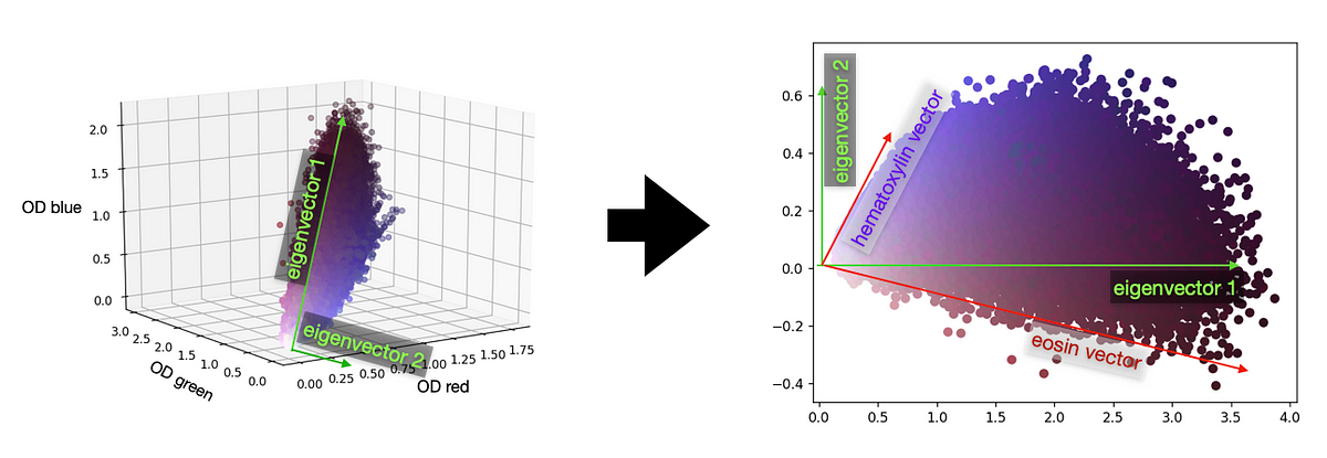
If you have seen a couple of microscopy images of tissue (histopathology images), you must have noticed that they come in all variants of colors. Even when the same dying chemicals (stains) are used, the visual appearance is influenced by so many factors that it can easily become a big problem if you work with those images algorithmically.The default stain in histopathology is a combination of two chemicals: hematoxylin and eosin. The first is responsible for the dark violet (or blueish) color of all acid components (like, e.g., the DNA, residing in the cellular nucleus), while the second is responsible for the pinkish color that gives structure to the surrounding tissue. It looks for example like this:
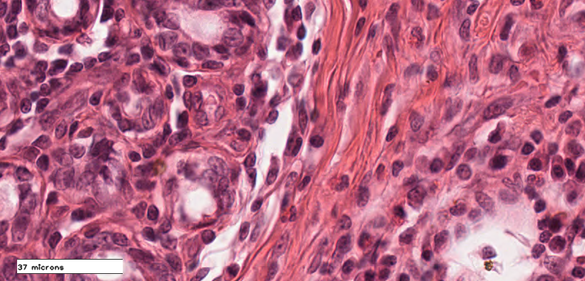
A histopathology image of breast tissue. Thanks to C. Bertram and R. Klopfleisch from FU Berlin for providing the whole slide image. Image by author.
I’ve already mentioned that the color of this varies widely across images. Let us briefly discuss why that is:One major role in this game is played by the staining chemicals themselves. Depending on the concentration, the duration of staining and even the temperature. To stabilize these conditions, automatic staining machines are used. To showcase the influence of duration the tissue resides in a stain, have a look at the next image:
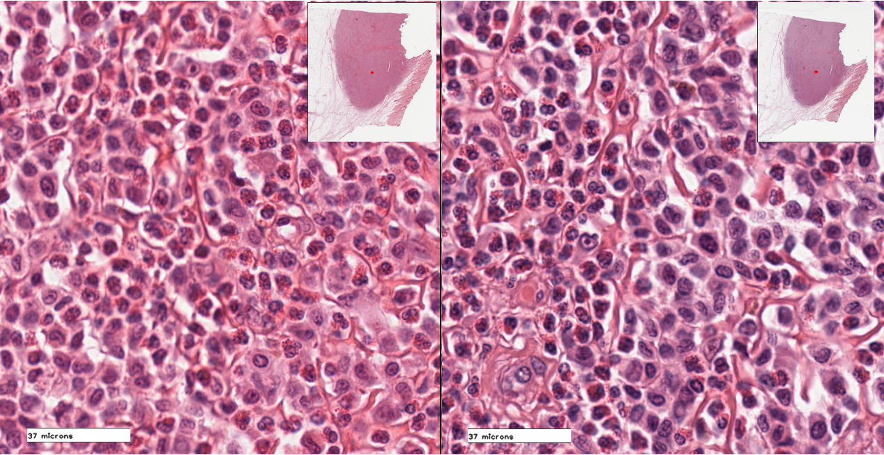
Histology image of canine cutaneous mast cell tumor, stained with H&E. Left image represents tissue left for 2 minutes in hematoxylin, while right side represents tissue left for 12 minutes in hematoxylin (Thanks to C. Bertram for providing the microscopy slides, image from author)
Both images represent slices of the exact same tumor, as can be seen in the overview thumbnail. However, the right image has a much stronger blue color component (caused by the much longer staining time in the hematoxylin solution).If we want to process these images, these variations often pose a problem. Especially when we have only a small number of cases, it is at times hard to get our recognition algorithms robust, especially if they have a high pattern recognition capacity, such as by using deep networks.
#deep-learning #normalization #data-science #microscopy #pathology #deep learning