This is part 2 of the application of Deep learning on X-Ray imaging. Here the focus will be on understanding X-ray images — with a special focus on Chest X-rays.
Interpreting Chest X-Rays:

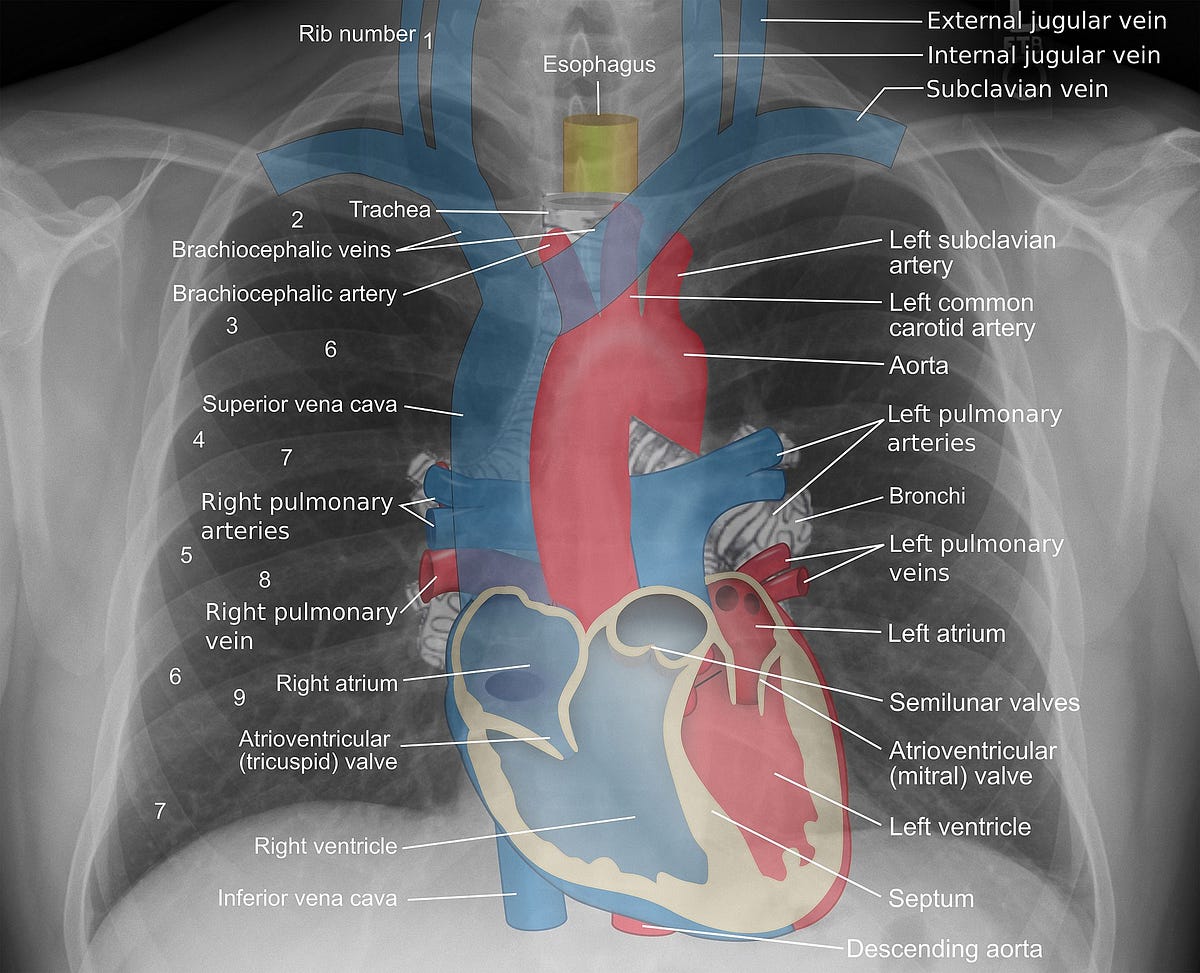
Figure 1. Chest X-Ray — 1) Lungs, 2) Right Hemidiaphragm, 3) Left Hemidiaphragm, 4) Right Atrium, 5) Left Atrium (By Diego Grez — Radiografía_pulmones_Francisca_Lorca.jpg, CC BY-SA 3.0, https://commons.wikimedia.org/w/index.php?curid=10302947. Editing by Author)
X-ray images are grayscale images, that is, the images have some pixels which are dark and some that are bright. In medical imaging terms, these are images that have values ranging from 0 to 255, where 0 corresponds to the completely dark pixels, and 255 corresponds to the completely white pixels.

Figure 2. The grayscale bar
Different values on the X-ray image correlate to different areas of density:
- Dark — Locations on the body which are filled with Air are going to appear black.
- Dark Grey — Subcutaneous tissues or fat
- Light Grey — Soft tissues like the heart and blood vessels
- Off White — Bones such as the ribs
- Bright White — Presence of metallic objects such as pacemakers or defibrillators
The way physicians interpret an image is by looking at the borders between the different densities. As in Figure 1, the ribs appear off-white because they are dense tissues, but since the lungs are filled with air, the lungs appear dark. Similarly below the lung is the hemidiaphragm, which is again a soft tissue and hence appears light grey. This helps us give a clear understanding of the location and extent of the lungs.
So, if two objects with different densities are present close to each other they can be demarcated in an X-ray image.
Now if something were to happen in the lungs, such as Pneumonia, then, the air dense lungs will change into water-dense lungs. This will cause the demarcation lines to fade since the pixel densities will start closing in on the grayscale bar.
For taking a chest X-ray, normally the patient is asked to stand, and the X-rays are shot from either front to back (Anterior-Posterior) or from back to front (Posterior-Anterior).
#artificial-intelligence #machine-learning #x-rays #deep-learning #deep learning