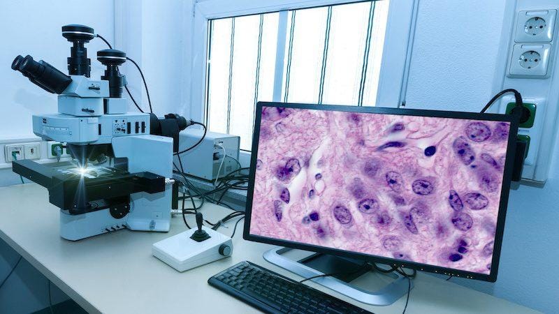
Histopathology is the study of diseases of tissues that involves the examination of microscopic slides consisting of tissues, cells, etc, that has been extensively used for the diagnosis of various forms of cancer. Histopathologists are medical experts who analyze cells or tissues under the microscope to make a diagnosis in order to come to a consensus regarding nature, the severity of diseases, and plan of action with regards to patient care.
With the advent of advanced equipment such as specialized scanning machines, strides in storage/cloud capabilities, it has now become quite easy to store Microscopic glass slides in the form of Digital slides on a computer for processing them. It has enabled remote diagnosis, faster analysis, and systematic and safe storage of pathology information. Recent events such as the global pandemic have shown us the importance of automating medical activities such as diagnostics. Not only will it help improve the accuracy and capacity, removing a lot of redundancy, but it will ensure less exposure for the Doctors and diagnosticians on the frontline.
Due to the rising applicability, scalability, and success of Artificial Intelligence and Machine Learning and its multidisciplinary nature, it is increasingly being applied to various fields. Medical Science is no different. A large number of procedures from Automated diagnosis to surgical procedures to drug discovery are leveraging Machine learning with promising results. Medical imaging for diagnosis using Machine Learning and Classical computer vision is a very fastly growing area of research.
Deep Learning, especially CNNs has become the methodology of choice when it comes to digital histopathology. This blog is a summarised review of the current state of research, the best practices, and the challenges in the field of Deep Learning for Histopathology. The review is structured based on the different deep learning approaches and discusses some of the popular, state of the art papers.
Challenges
Unlike normal image data used for General computer vision tasks, dealing with Whole slide Digital pathology images poses a unique set of challenges.
The most significant challenge is the size of Whole slide images. These slides are usually gigabytes in size and thus pose a computation bottleneck both in terms of storage, processing, and compatibility with deep learning algorithms. A large percentage of literature dealing with deep learning deals with this problem using a Patch extraction/tiling method. They mostly use a sliding window-based approach to extract smaller patches which are then sent for further processing. Another common approach is to downsample the whole slide images to a tractable size but this leads to information loss, which in turn affects the performance of deep learning algorithms.
Researchers have begun to notice limiting flaws in the patch extraction approach too.
- To make sure important information is not lost, significant attention needs to be given to ensure maximum overlap among patches by carefully adjusting the strides
- In order to perform any classification/prediction at the whole slide image level, we need to go through the entire slide, and hence a large number of patches need to be processed which makes the training and prediction process tedious and computationally expensive.
- When we deal with patches we are dealing with a very small region at a time, hence a lot of global contextual information is lost which limits the ceiling performance.
Overcoming these limitations of the patch extraction method on whole slide digital pathology image analysis using deep learning has become a hot topic of research on its own. A number of solutions have been proposed and experimented with.
One of the significant viable solutions proposed is to use Sparse Coding of pathology slides for learning features and inferring representations of Cancer histology slides.
LSTMs and Conditional Random Fields are increasingly being used in combination with CNNs to model the correlation between neighboring patches, in order to capture larger contextual information.
Cascaded CNNs and Visual attention-based methods are being utilized to enforce a region selection method so that only the most relevant, diagnostically useful areas of the whole slide image is used for prediction and training.
A highly ingenious solution called slide graph¹ proposed in a work presented very recently in CVPR’20 addresses the issue of size by using a Graph Convolutional neural network-based model.
#machine-learning #computer-vision #medical-imaging #histopathology #deep-learning #deep learning